Table of Contents >> Show >> Hide
- What Is an Oral Cholecystogram?
- Purpose: Why Would a Doctor Order an Oral Cholecystogram?
- Who Should Not Have an Oral Cholecystogram?
- Preparation: How to Get Ready (Without Overthinking It)
- Procedure: What Happens During an Oral Cholecystogram?
- Does It Hurt? What Does It Feel Like?
- Results: What Can an Oral Cholecystogram Show?
- Risks and Side Effects
- Alternatives to an Oral Cholecystogram (What You’re More Likely to See Today)
- FAQ: Quick Answers to Common Questions
- Experiences & Practical Tips (The “What It’s Actually Like” Section)
- Conclusion
- SEO Tags
If your gallbladder could talk, it would probably say, “I’m small, I’m moody, and when I’m upset, I make the whole
upper-right side of your belly file a complaint.” An oral cholecystogram (also called
oral cholecystography or OCG) is one of the classic ways doctors used to “see”
the gallbladder on X-rayby having you swallow special iodine-based contrast tablets the night before.
These days, OCG is much less common because ultrasound, CT, MRCP, and nuclear medicine tests like the
HIDA scan usually answer the same questions faster and with fewer prep steps. Still, you may run into an oral
cholecystogram in certain clinics, older protocols, or special situations. If your provider ordered one, don’t panic:
it’s generally straightforwardjust a little “retro,” like a flip phone that still somehow works.
What Is an Oral Cholecystogram?
An oral cholecystogram is an imaging test that uses X-rays to evaluate the gallbladder after you take
an oral contrast agent. Here’s the basic plot:
- You swallow contrast tablets (typically iodinated) the evening before your imaging.
- Your intestines absorb the contrast, your liver processes it, and it’s excreted into bile.
- The contrast collects in your gallbladder, helping it “light up” on X-ray.
- Radiology takes images to look for gallstones, abnormal shape, poor emptying, or blockage.
A key detail: many gallstones (especially cholesterol stones) don’t show up well on a regular plain X-ray. In an oral
cholecystogram, stones may appear as filling defects inside an otherwise contrast-filled gallbladder.
Purpose: Why Would a Doctor Order an Oral Cholecystogram?
The test is designed to evaluate gallbladder structure and functionparticularly when gallbladder disease is suspected.
Common reasons include:
1) Suspected Gallstones (Cholelithiasis)
If symptoms point to gallstoneslike episodes of right upper quadrant pain after fatty meals, nausea, or “biliary colic”
imaging helps confirm the diagnosis. OCG can detect gallstones as defects within the contrast-filled gallbladder and can
sometimes reveal polyps or masses.
2) Possible Inflammation (Cholecystitis) or Blockage
A gallstone can obstruct the cystic duct, triggering inflammation. One classic “red flag” pattern in OCG is a
non-visualized gallbladder (meaning the gallbladder doesn’t fill with contrast as expected). That can
happen if the cystic duct is blocked or if the gallbladder isn’t functioning well.
3) Functional Clues (How Well the Gallbladder Concentrates and Empties)
Some protocols include images after a fatty snack or drink to stimulate gallbladder contraction.
In simple terms: does the gallbladder empty like it should, or does it behave like it’s “on strike”?
Why It’s Rare Today
Modern imaging is often quicker, easier to schedule, and more informative:
ultrasound is commonly first-line for gallstones, HIDA scan is excellent for
functional obstruction and acute cholecystitis questions, and MRCP can map bile ducts without
iodinated oral contrast. But if OCG is what’s on the menu, it can still provide useful information.
Who Should Not Have an Oral Cholecystogram?
Your clinician will weigh risks and choose the right test. OCG may be avoided or used with extra caution if you have:
- Known allergy to iodinated contrast agents (or a history of significant reactions).
- Pregnancy (X-rays are generally avoided unless truly needed).
- Severe liver disease or significant jaundice (the contrast may not be processed/excreted as needed).
- Severe vomiting or inability to keep tablets down.
- Certain bowel issues or recent GI contrast studies that can obscure images (your team will advise).
Always tell your provider about allergies, pregnancy status, and major liver/kidney conditions. This is not the moment
for “I’m sure it’s fine” optimismsave that energy for your fantasy football lineup.
Preparation: How to Get Ready (Without Overthinking It)
Prep matters because the contrast needs to be absorbed, processed by your liver, and concentrated in the gallbladder.
Different facilities have slightly different instructions, but this is the common playbook:
1) Diet the Day Before
Many protocols recommend a low-fat or fat-free diet the day before. Why? Too much fat can stimulate
gallbladder contraction at the wrong time and mess with how well the gallbladder concentrates contrast.
Practical low-fat options: lean chicken or fish, vegetables, fruit, bread, rice, broth-based soups, skim milk (if allowed).
Translation: it’s not a “foodie” day. It’s a “functional adult” day.
2) Taking the Contrast Tablets
You’ll take the prescribed contrast pills the evening before the exam. Some instructions use a series like
six tablets with specific timing (for example, spaced out over hours or taken one every few minutes),
and your facility will tell you the exact schedule. Follow it preciselythis is not a “close enough” situation.
3) Fasting the Morning of the Exam
Most instructions require no food or drink the morning of the procedure (or only minimal sips for
necessary medicationsask your provider).
4) Sometimes: Bowel Prep (Laxative or Enema)
If you’ve had recent GI imaging with contrast (like a barium study) or if your facility wants a clearer view, you might
be instructed to take a laxative or enema to reduce bowel contents that can obscure the gallbladder region on X-ray.
Prep Checklist (Quick Version)
- Confirm timing for your contrast tablets.
- Go low-fat the day before (unless told otherwise).
- Fast the morning of the test.
- Bring your allergy history and medication list.
- Wear comfortable clothing (and consider leaving “tight waistband day” for another time).
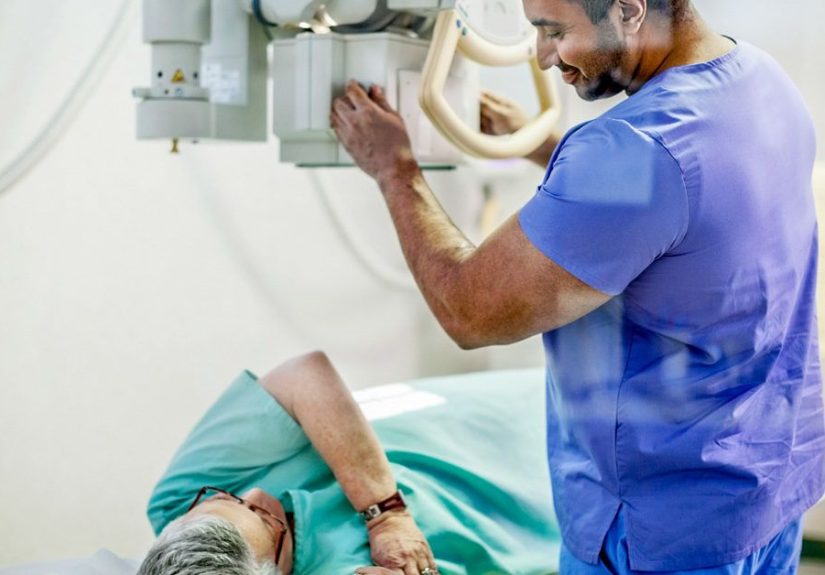
Procedure: What Happens During an Oral Cholecystogram?
The imaging portion is usually outpatient and doesn’t involve needles for the OCG itself. Here’s what to expect:
Step 1: Check-In and Safety Questions
You’ll confirm your identity, review allergies (especially to contrast), and answer pregnancy screening questions when relevant.
Step 2: Abdominal X-Rays (Sometimes with Fluoroscopy)
A technologist takes several abdominal images while you lie on an exam table (and sometimes while you stand).
You may be asked to change positions so the gallbladder region can be seen clearly.
Step 3: Sometimes a Fatty Meal Challenge
In some protocols, you’ll be given a fatty snack or drink after initial images. Fat triggers the
gallbladder to contract. Follow-up images can show how the gallbladder empties and whether contrast moves into the
bile ducts and intestine.
Step 4: More Images (and Possibly a Repeat Day)
If the gallbladder doesn’t show up well, your provider might recommend repeating the study the next day. This doesn’t
automatically mean “bad news”it can reflect timing, absorption, or other factorsthough it can also signal obstruction
or poor gallbladder function.
Does It Hurt? What Does It Feel Like?
The imaging itself is typically painless. The most common “sensations” are boredom and mild annoyance at being asked to
hold still right when your nose itches.
Side effects are more likely from the oral contrast tablets than the X-rays. Some people experience:
- Mild nausea
- Diarrhea or loose stools
- Abdominal cramping
Results: What Can an Oral Cholecystogram Show?
Your images are interpreted by a radiologist, and a report is sent to the ordering clinician. While every case is unique,
here are common patterns:
1) Normal Gallbladder Visualization
Contrast fills the gallbladder normally, suggesting that absorption, liver processing, bile excretion, and cystic duct
flow are functioning well.
2) Filling Defects (Possible Gallstones or Polyps)
If the gallbladder is outlined by contrast but has dark “holes” or defects, that can suggest gallstones or sometimes polyps.
Your provider may correlate this with symptoms, labs, and other imaging.
3) Non-Visualized Gallbladder
If the gallbladder does not fill with contrast, possibilities include cystic duct obstruction (often from a stone),
significant inflammation, or reduced function. This finding often prompts follow-up evaluation with modern tests like
ultrasound or a HIDA scan.
4) Poor Emptying After Fatty Meal
If a fatty meal is used and the gallbladder doesn’t contract as expected, that may suggest impaired function. The next
steps depend on symptoms and the broader clinical picture.
Risks and Side Effects
Oral cholecystography is generally safe, but like any medical test, it has trade-offs:
Contrast-Related Effects
- Diarrhea, nausea, vomiting, stomach cramps
- Allergic reactions ranging from rash/itching to more serious reactions (rare)
- In some cases, the test may not work well if the contrast doesn’t reach the gallbladder
X-Ray Exposure
The test uses ionizing radiation. The dose is typically low, but it’s still a factorone reason providers often choose
ultrasound first. If you’re pregnant or might be, tell your care team before imaging.
Alternatives to an Oral Cholecystogram (What You’re More Likely to See Today)
If you’re reading this and thinking, “Why haven’t I heard of this test?”that’s because modern gallbladder imaging often
uses other tools:
Abdominal Ultrasound
Common first-line test for gallstones. It’s quick, doesn’t use radiation, and is widely available.
HIDA Scan (Hepatobiliary Scan)
A nuclear medicine test that evaluates bile flow and gallbladder function using an injected tracer. It’s especially useful
when acute cholecystitis or functional obstruction is suspected.
CT Scan
Helpful for complications and broader abdominal evaluation (though it can miss some gallstones).
MRCP (Magnetic Resonance Cholangiopancreatography)
MRI-based imaging that maps the bile ducts and pancreatic duct without invasive dye injection.
ERCP (Endoscopic Retrograde Cholangiopancreatography)
A scope-based procedure that can diagnose and treat certain bile duct problems (like removing duct stones), but it’s more
invasive than imaging-only tests.
FAQ: Quick Answers to Common Questions
How long does an oral cholecystogram take?
The prep takes the longest (taking tablets the evening before). The imaging appointment itself is usually much
shorteroften under an hourdepending on the protocol and whether post-meal images are needed.
Can I drive myself home?
Usually yes, because sedation is not typically involved. If your facility pairs it with other tests or gives special meds,
follow their instructions.
What if I throw up the tablets?
Call your provider. If you can’t keep the contrast down, the test may need to be rescheduled or replaced with another
imaging option.
Why would my gallbladder not show up?
Non-visualization can occur if the contrast doesn’t reach the gallbladder or if gallbladder function/duct flow is impaired.
Your clinician will interpret this in context and may recommend ultrasound or a HIDA scan next.
Experiences & Practical Tips (The “What It’s Actually Like” Section)
Let’s talk about the part patients care about most: the lived reality of an oral cholecystogram. Not the textbook version,
but the “How do I get through this without accidentally sabotaging the test?” version.
The night-before routine is the main event. People often expect the imaging appointment to be the hard
part, but the real challenge is remembering to take the contrast tablets exactly as instructed. The simplest hack is to
set alarms on your phone with labels like “Gallbladder Glitter Pill #1” (or something less dramatic if you’re a serious
person). Missing a dose or taking them at the wrong intervals can reduce how well your gallbladder fills and may lead to
a repeat studynobody wants a sequel.
The low-fat diet feels suspiciously healthy. Many patients report that the day-before menu is the weirdest
part emotionally: you’re preparing for a gallbladder test, and suddenly your kitchen looks like a wellness influencer’s
story highlight. Keep it simplelean protein, rice, fruit, vegetables, broth soups. It’s not forever; it’s a one-day
temporary truce with fried foods.
GI side effects are the most common “plot twist.” The contrast tablets can cause diarrhea, nausea, or
cramping for some people. Most describe it as mild and temporarybut it’s wise to plan accordingly. If you’re taking the
tablets at night, staying near a bathroom isn’t “being dramatic,” it’s being strategic. Hydrate as allowed by your
instructions, and don’t stack other stomach-irritating choices (like a late-night spicy feast) on top of the contrast.
Fasting is manageableuntil your brain starts negotiating. On the morning of the test, patients often
say they’re surprised by how quickly they start rationalizing snacks: “Technically, is coffee a food?” (No. Also: ask
your facility before taking anything.) If you’re someone who wakes up hungry, plan distractionswork, a walk, a podcast,
anything that keeps you from staring at your toaster like it betrayed you.
In the imaging room, the vibe is calm and procedural. Technologists guide you through positionsturning
slightly, lying still, maybe standing for a view. Patients often say it feels like a short photo shoot where the camera
is a fluoroscope and the theme is “upper abdomen, natural lighting.” The only “skill” required is holding still and
following directions.
The fatty meal part can feel oddly anticlimactic. If your protocol includes a fatty snack or drink to make
the gallbladder contract, some people expect fireworks. Usually it’s just: you consume the item, wait, and take more
images. If you’re sensitive to fat or already symptomatic, tell the staffyour team can adapt or choose alternatives.
Results anxiety is realso keep expectations grounded. A “normal” OCG doesn’t magically explain every
stomach ache, and an “abnormal” one doesn’t automatically mean surgery tomorrow. Many patients feel relief when they
remember the goal: gather clues, then decide the next step with a clinician who can interpret the full storysymptoms,
labs, physical exam, and imaging together.
Pro-tip: treat the instructions like a recipe, not a suggestion. The test depends on timing, absorption,
and gallbladder filling. If you want the most accurate result (and the lowest chance of repeating it), follow the prep
steps the way you’d follow a baking recipe for a fancy cake. Eyeballing works for chili; it’s not ideal for diagnostic imaging.
Conclusion
An oral cholecystogram is an old-school-but-still-valid way to evaluate the gallbladder using X-rays after
you take contrast tablets the night before. It can help detect gallstones, suggest obstruction or reduced
function, and guide next stepsespecially when paired with clinical symptoms like biliary colic or suspected cholecystitis.
While modern tests (ultrasound, HIDA scan, MRCP, CT) are more common today, OCG remains a useful piece of the diagnostic
toolbox in select settings.
If you’re scheduled for one, the best thing you can do is simple: follow the prep instructions closely, report allergies
and pregnancy status, and let your care team connect the dots afterward.