Table of Contents >> Show >> Hide
- What Is a Knee MRI Scan?
- Purpose: Why Doctors Order a Knee MRI
- What a Knee MRI Can Show
- Knee MRI vs. X-ray, Ultrasound, and CT
- How to Prepare for a Knee MRI
- Procedure: What Happens During a Knee MRI
- Will I Need Contrast for a Knee MRI?
- After the Scan: Results and Next Steps
- Risks of a Knee MRI Scan
- How to Make a Knee MRI Easier (Practical Tips)
- FAQs
- Conclusion
- Real-World Experiences (What People Commonly Report)
A knee can be dramatic. One minute you’re walking like a normal human, the next you’re negotiating stairs like they’re
a hostile mountain range. When knee pain, swelling, or a suspicious “pop” refuses to mind its business, your
healthcare provider may order a knee MRI scan to find out what’s happening inside the jointwithout
guessing, squinting, or consulting a magic 8-ball.
This guide breaks down what a knee MRI is for, what the scan is like from start to finish, and the real-world risks
(spoiler: the loud noises are normal, and the magnet is not here to make friends with your metal).
What Is a Knee MRI Scan?
MRI stands for magnetic resonance imaging. Unlike X-rays or CT scans, MRI doesn’t use
ionizing radiation. Instead, it uses a strong magnetic field and radio waves to create detailed imagesespecially of
soft tissues like ligaments, tendons, cartilage, and menisci. That’s why knee MRI is so useful: most
knee problems aren’t “bone-only” issues.
Purpose: Why Doctors Order a Knee MRI
A knee MRI is usually ordered when your symptoms suggest a problem that’s hard to confirm with a physical exam alone
or with basic imaging like X-rays. X-rays are great for bones, but knee pain often comes from the “support staff”
inside the joint.
Common reasons for a knee MRI
- Suspected meniscus tear after a twist, pivot, or sports injury
- Ligament injuries (ACL, PCL, MCL, LCL)especially after a sudden stop or awkward landing
- Cartilage damage or early joint wear that doesn’t show well on X-ray
- Tendon problems (like patellar tendon or quadriceps tendon issues)
- Persistent swelling, fluid buildup (effusion), or suspected inflammation
- Unexplained pain that isn’t improving with rest, therapy, or time
- Suspected infection or unusual bone marrow changes
- Cysts (like a Baker’s cyst behind the knee)
- Tumors or masses (rare, but MRI is excellent for characterizing them)
- Pre-surgical planning or evaluation after knee surgery
Example: If you have pain along the joint line, swelling after activity, and “catching” or locking, your provider may
suspect a meniscus tear. MRI is often considered the preferred imaging option for diagnosing acute meniscus tears
because of its accuracy.
What a Knee MRI Can Show
Think of your knee as a busy intersection of bones, cartilage, ligaments, tendons, and shock absorbers. MRI can show
details that are simply invisible on many other tests.
Key structures MRI evaluates
- Menisci (medial and lateral): tears, degeneration, displaced fragments
- Ligaments: sprains, partial tears, full ruptures
- Cartilage: thinning, defects, wear patterns tied to arthritis
- Tendons: tendinitis, partial tearing
- Bone marrow: bruises, stress injuries, subtle fractures
- Synovium and fluid: inflammation, effusions, synovitis
- Soft tissue masses: cysts or other abnormal growths
Knee MRI vs. X-ray, Ultrasound, and CT
Imaging isn’t a popularity contestit’s “best tool for the job.”
- X-ray: Best for fractures, alignment, and moderate-to-advanced arthritis changes.
- Ultrasound: Useful for some tendon issues, fluid collections, and guided injections; not as detailed for deep joint structures.
- CT: Great for complex bone detail; sometimes used if MRI isn’t possible.
- MRI: Best all-around view for soft tissues and many internal knee injuries.
How to Prepare for a Knee MRI
Preparation is usually simple, but the safety screening matters. MRI uses a powerful magnet, so your imaging team will
ask about implants, devices, or metal in or on your body.
What to do before you arrive
- Know your medical history: pacemakers, aneurysm clips, cochlear implants, metal fragments, implanted pumps, or prior surgeries.
- Dress simply (or expect a gown): avoid metal zippers, snaps, underwire, and metallic threads.
- Leave valuables at home: jewelry, watches, and cards are better off not meeting a high-powered magnet.
- Tell them about claustrophobia ahead of time: options may include coaching, music, open MRI (in some centers), or medication.
- Ask about eating and drinking rules: many MRIs don’t require fasting, but some centers may give specific instructionsespecially if sedation or contrast is planned.
If you’ve ever worked around metal grinding (welding, machining), mention it. In certain situations, providers may
check for metal fragmentsespecially near the eyesbecause the magnet can be dangerous if metal is present.
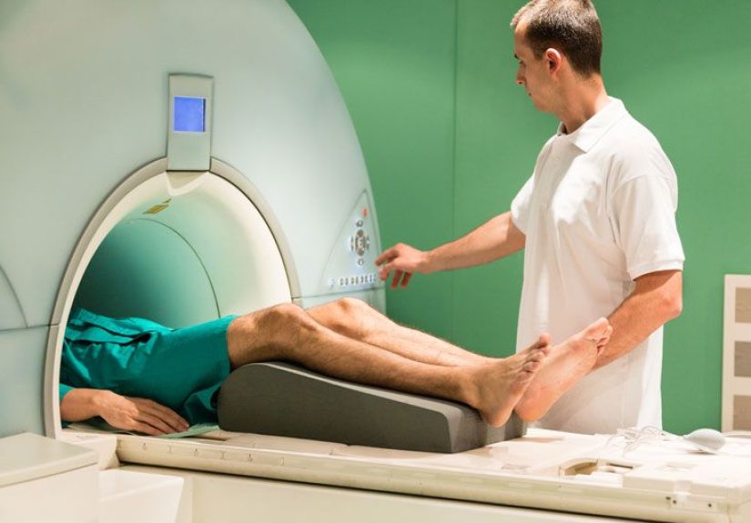
Procedure: What Happens During a Knee MRI
Step 1: Check-in and safety screening
You’ll complete a screening form and go over implants or prior surgeries. This isn’t paperwork for paperwork’s sake
it helps the team confirm MRI is safe for you and prevents last-minute cancellations.
Step 2: Changing and setup
You’ll be asked to remove metal items and may change into a gown. Then you’ll lie on the MRI table. For a knee MRI,
the technologist positions your knee and may use a supportive device or padding to keep you comfortable and still.
Step 3: The scan itself (aka “The Very Loud Donut Experience”)
The table slides into the MRI machine. The scanner makes loud tapping or knocking sounds during image sequences.
You’ll likely get ear protection and sometimes headphones. The technologist can usually see and hear you, and you may
have a call button.
Step 4: Staying still
Stillness is everything. Even small movements can blur the images and force repeat sequences. If you’re in pain, tell
the team before the scan startssometimes they can adjust positioning or support to help you hold still.
How long does a knee MRI take?
Many knee MRIs take roughly 20 to 45 minutes, but timing varies depending on the protocol, whether
contrast is used, and whether additional sequences are needed.
Will I Need Contrast for a Knee MRI?
Not always. Many knee MRIs are done without contrast, especially for common sports injuries like ACL
or meniscus tears. But contrast can help in certain situations, such as evaluating:
- infection or inflammatory conditions
- tumors or unusual masses
- post-surgical scarring vs. recurrent injury
- specific cartilage or joint lining concerns
What is MRI contrast?
MRI contrast is often a gadolinium-based agent given through an IV. It can make certain tissues and
blood flow patterns easier to see.
What is an MR arthrogram?
In selected cases, your provider may order an MR arthrogram, where contrast is injected
into the knee joint (not just into a vein) before the MRI. This can help highlight internal joint structures
in more detail. It involves a needle and sometimes imaging guidance to place the contrast accurately.
After the Scan: Results and Next Steps
After your MRI, you can usually return to normal activities right away unless you received sedation. A radiologist
interprets the images and sends a report to the clinician who ordered the study.
What your report might mention
- Meniscus: tear type, location, displacement
- Ligaments: sprain vs. partial vs. full tear
- Cartilage: thinning, defects, arthritis-related changes
- Bone: bruising, stress injury, fracture signs
- Fluid/inflammation: effusion, synovitis, cysts
Tip: MRI findings should match your symptoms. Plenty of people have “abnormal” findings that aren’t causing pain.
Your clinician will interpret results in context, not as a standalone plot twist.
Risks of a Knee MRI Scan
For most people, MRI is considered very safe. Still, “safe” doesn’t mean “zero things to know about.” Here are the
real risks and considerations.
1) Metal and implanted devices
The strongest risk category is magnet + metal. Some implanted devices can malfunction, heat up, move,
or distort images. That’s why screening is strict. Many modern implants are MRI-conditional (safe under specific
conditions), but the team needs details.
2) Claustrophobia and anxiety
If enclosed spaces make your brain hit the panic button, you’re not alone. Options may include coaching, breathing
strategies, music, having the technologist talk you through sequences, or medication. Some facilities offer open MRI,
though image quality can vary.
3) Noise and discomfort
MRI scanners can be loud. Ear protection helps, but don’t be surprised if it sounds like a robot building a deck in
the next room. Discomfort can also come from holding still or keeping the knee in one position.
4) Sedation risks (if used)
Some patientsespecially children or people with severe anxietymay receive sedation. Sedation is generally safe when
monitored, but it carries risks like over-sedation and requires follow-up instructions (like not driving afterward).
5) Contrast risks (if contrast is used)
Most people tolerate gadolinium contrast well. Possible issues include:
- Allergic-like reactions: uncommon and usually mild (for example, hives or itching). Severe reactions are rare.
-
Kidney disease considerations: older gadolinium agents were linked to a rare condition called
nephrogenic systemic fibrosis (NSF) in people with severe kidney failure. Newer agents are considered much safer, but your team may check kidney function if you’re at risk. - Gadolinium retention: small amounts can remain in the body for months to years; regulators have noted no clear harmful effects identified to date in patients with normal kidney function, but class warnings and patient education exist.
6) Pregnancy considerations
MRI is often used when medically necessary, but contrast is typically avoided during pregnancy unless it’s essential.
If you’re pregnant or might be, tell your provider and the imaging center before the scan.
How to Make a Knee MRI Easier (Practical Tips)
- Bring your implant info (device card, surgery details) if you have any.
- Ask for comfort supports: pillows, padding, and positioning adjustments can help you stay still.
- Plan for the noise: earplugs/headphones are normaluse them.
- If you’re anxious, say it early: the earlier you mention claustrophobia, the more options you’ll have.
- Don’t “tough it out” in silence: tell the technologist if pain is making it hard to hold still.
FAQs
Does a knee MRI hurt?
The scan itself shouldn’t hurt. The most common “ouch” is positionalholding still or keeping the knee in one position.
If you’re already in pain, positioning can be uncomfortable, but the team can often adjust supports.
Can I drive myself home?
Yes, if you did not receive sedation. If you took medication for anxiety or were sedated, you’ll need a ride.
What if I have a knee replacement or screws?
Many orthopedic implants are compatible with MRI under certain conditions, but they can sometimes distort images.
Always disclose implants so the facility can confirm safety and choose the best imaging approach.
How soon will I get results?
It depends on the facility and urgency. Some results come back within a day or two; others may take longer. Ask your
ordering provider what to expect and how results will be communicated.
Conclusion
A knee MRI scan is one of the most informative tools for diagnosing internal knee problemsfrom
ligament injuries and meniscus tears to cartilage damage and inflammation. The procedure is noninvasive, typically
doesn’t require special preparation, and is considered very safe for most people. The biggest “risks” are really
screening issues (metal and implants), comfort issues (noise and stillness), and
contrast considerations for people with certain kidney conditions.
If you’re scheduled for a knee MRI, the best move is simple: be honest about implants, speak up about anxiety or pain,
and show up ready to hold still like you’re winning the world’s quietest contest. Your kneeand your future self on
the stairswill thank you.
Real-World Experiences (What People Commonly Report)
Let’s talk about the part most brochures gloss over: what a knee MRI feels like in real life. Not “feel” as
in pain (usually none), but the whole vibebecause the vibe matters when you’re lying still inside a very expensive
magnet that sounds like it’s remixing construction noises.
Experience #1: The weekend athlete with a twist injury. A common story: someone pivots during
basketball, soccer, or a perfectly innocent attempt at “just jogging,” then hears or feels a pop. The knee swells
later, and walking feels unstable. Many people describe arriving for the MRI with two emotions: curiosity and
suspicioncuriosity about what’s wrong, and suspicion that the machine is going to be terrifying. Usually, the
surprise is how routine it is. The technologist positions the leg, explains the sounds, and suddenly the main job is
staying still. People often say the hardest part isn’t fearit’s resisting the urge to adjust their foot or scratch an
itch when the scan is mid-sequence. (Your nose may randomly itch. This is a scientific law.)
Experience #2: The chronic knee pain mystery. Another frequent scenario is a person with months of
pain that doesn’t match their X-ray. They may have trouble with stairs, squats, kneeling, or long walks. For these
patients, the MRI experience is often less about “oh no, I’m injured” and more about “please let this explain why my
knee hates me.” Many report feeling relief just having a planbecause even if the MRI shows arthritis changes,
cartilage wear, or inflammation, it turns vague pain into specific next steps (physical therapy focus, injections,
training modifications, or surgical conversations when appropriate).
Experience #3: Claustrophobia and the ‘talk-me-through-it’ approach. People who are anxious about
enclosed spaces often do best when they tell the imaging center early. Many say it helps when the technologist gives a
clear timeline (“This next set is 3 minutes; you’re doing great”), offers music, and reminds them they can communicate
at any time. Some patients practice a simple strategy: eyes closed before entering the scanner, slow breathing, and
mentally “chunking” the scan into short segments. Others request medication for anxiety ahead of time and emphasize
that it transforms the experience from white-knuckle to manageable. The consistent theme? The scan is easier when you
treat it like a series of short sprints rather than one long endurance event.
Experience #4: Contrast worries (and what it’s usually like). When contrast is used, people commonly
report that the IV part is the most noticeable moment. The injection itself is typically quick, and most people feel
normal afterward. Some people go in worried about “dye reactions” because they’ve heard stories about contrast with
other scans. In practice, most describe it as anticlimacticthough anyone with kidney disease concerns, prior contrast
reactions, or multiple past MRIs tends to appreciate a clear explanation of why contrast is (or isn’t) needed this
time.
Experience #5: Getting the report back. A very human reaction is immediately Googling phrases from the
report. (We’ve all done it. The internet has opinions.) Many people find it more helpful to discuss the results with a
clinician who can translate radiology language into “what this means for your life.” For example, a meniscus tear on
MRI doesn’t automatically mean surgerysymptoms, tear type, activity level, and overall knee health matter. Patients
often say the best outcome of the MRI isn’t just the imagesit’s clarity, direction, and fewer “what ifs.”
Bottom line: most knee MRI experiences are described as inconvenient but manageablemore “long and noisy” than
“scary and painful.” If you prepare for stillness, communicate openly, and show up with zero metal, you’re already
doing it right.